The elbow complex consists of the compound elbow joint and the radioulnar joints, with all three joint articulations found within the anterior joint capsule. The elbow joint articulation is technically classified as a trochoginglymoid joint. A description of the three joint articulations is detailed below:

- Humeroulnar joint (HUJ) – this is a (modified) hinged (ginglymus) joint that provides flexion / extension motions. The surface-on-suface motion is predominantly gliding with the concave surface of the ulna (trochlea notch) glides over the convex surface humerus (trochlea) and the concave radial head gliding along the capitulum of the humerus. During flexion the elbow joint exhibits anterior glide, whereas during extension the elbow joint exhibits posterior glide. This joint provides most of the structural stability to the elbow through inherent osseous constructs of the humerus and ulna.


- Humeroradial joint (HRJ) – this is a ball and socket joint involved in elbow flexion/extension as well as forearm pronation/supination; permits continuous contact between the radial head and the capitulum during pronation and supination, as radius spins about its own axis.
- Proximal Radioulnar joint (RUJ) – this is a pivot (trochoid) joint in which the head of the radius articulates with the radial notch of the ulna, and is involved in only forearm pronation/supination. There is also a distal RUJ joint that is outside of the joint capsule. The two RUJ joints function as one joint.


Despite having three distinct joint articulations in the elbow joint capsule, the elbow complex only has two degrees of freedom (DOF): flexion/extension along the HUJ hinge joint and pronation/supination along the RUJ pivot joint. The HRJ joint is in very close proximity to both the HUJ and proximal RUJ which restricts the joint motion from three to two DOF. Due to the close coupling of the two DOF motions in the elbow, the motions are not considered as individual joints, but rather biomechanically as cardinal joint motion.
The cubitus angle is more commonly referred to as the carrying angle which describes the valgus angle of the forearm as it projects laterally relative to the humerus when the elbow is fully extended and the forearm is fully supinated. In men, the average carrying angle is 11 to 14 degrees. In females, the average carrying angle is 14 to 15 degrees. The carrying angle is approximately 1 degree larger in the dominant arm as well. The carrying angle decreases as the elbow is flexed and when the forearm is pronated, the carrying angle disappears.
To really understand how the elbow moves, we need to understand what muscle groups are involved. Elbow rotations are fairly complex given three bones, including two bones in the forearm, and 9 different muscular attachments, which can be classified by 4 different muscle groups:
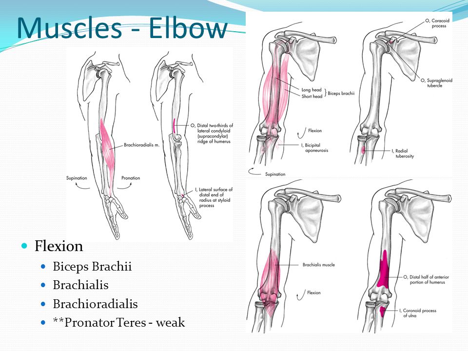
- Elbow flexors
- Brachialis – a single joint muscle that is a major flexor of the elbow. Arises from the entire anterior surface of the distal humerus and inserts on the coronoid process of the ulna. Some of the fibers insert into the anterior joint capsule and are thought to help retract the capsule during flexion.
- Biceps brachii – major flexor of the elbow where long head arises from a tubercle above the glenoid cavity of the scapula, travels through the shoulder joint between the greater and lesser tubercles, and along the bicipital groove before it merges with the body. The short head arises from the coracoid process of the scapula, and the muscular fibers join with those of the long head about halfway down the humerus. The tendon of the biceps inserts at the posterior aspect of the radial tuberosity, which means the biceps brachii is a powerful supinator as well. Long head is bi-articular so force production is dependent upon shoulder position. The function of the bicep changes with pronation, which is easily observed by flexing the elbow 90 degrees and rotating the forearm from full pronation into full supination while feeling the biceps bulge.
- Brachioradialis – originates from a lengthy lateral ridge on the mid to distal humerus and inserts via a long tendon on the base of the radial styloid process. Activates when the forearm is pronated and joint stability is needed. Active in flexion of the elbow and in also aids in rapid extension where it counters the centrifugal force produced by the movement.
- Pronator teres – assists only slightly in elbow flexion, whereas main function is to assist in pronation.

- Elbow extensors
- Triceps brachii – principal extensor of the elbow due to the massive tendon of insertion onto the olecranon of the ulna. Long head originates from a tubercle below the glenoid cavity. Long head is bi-articular and thus force production is dependent upon shoulder position. Lateral head originates from the lateral posterosuperior shaft of the humerus. Medial, or deep, head originates from the posteroinferior humerus and is covered up by the body of the muscle. Both medial and lateral heads are single joint muscles.
- Anconeus – much smaller muscle, that runs from the lateral epicondyle to the lateral olecranon and superior ulna. Assists the triceps in extension and also plays a small role in pronation. Helps to stablize the elbow during forearm pronation. Acts against valgus stress and helps to clear the joint space of soft tissue to permit full elbow extension.

- Forearm pronators
- Pronator quadratus – a square shaped muscle running between the anterior surfaces of the distal ulna and radius. This muscle offers superior mechanical advantage when it contracts, as it pulls the radius across the ulna in pronation.
- Pronator teres – originates from the medial epicondyle of the humerus and coronoid process of the ulna, and inserts on the midlateral surface of the radius. This is a major pronator of the forearm and assists in flexion of the elbow as well. Is active during fast pronation or pronation against resistance. Also acts to stabilize proximal RUJ.
- Brachioradialis – primarily an elbow flexor, but it can assist in the initial stages of pronation from a supinated position. It also works the opposite way as well, and helps to move the radius to a position intermediate between full pronation and full supination.

- Forearm supinators
- Biceps brachii – the more powerful supinator of the elbow, it tends to “uncross” the upper radius from a pronated position due to its insertion on the posterior aspect of the radial tuberosity.
- Supinator – originates in two layers. The superficial layer runs from the lateral epicondyle of the humerus, whereas the deep layer runs from the supinator ridge located just below the posterior radial notch of the ulna. Supinator wraps around the radius and inserts between the neck and insertion of the pronator teres. It helps to maintain supination with the biceps brachii by returning the proximal radius from a pronated position to the anatomical position of forearm supination.


